Why MFM Specialists Matter: What Every Expecting Mother Should Know Before Antenatal Care

Specialists in Maternal-Fetal Medicine (MFM) play a vital role in managing high-risk pregnancies. With advanced technology, structured care planning, and a multidisciplinary approach, they help reduce risks for both mother and baby from pregnancy through delivery.
Importance of Maternal-Fetal Medicine (MFM)
MFM plays a crucial role in helping high-risk pregnant women achieve safer pregnancies and deliveries by focusing on complication prevention, accurate diagnosis, and personalized care planning tailored to each individual.
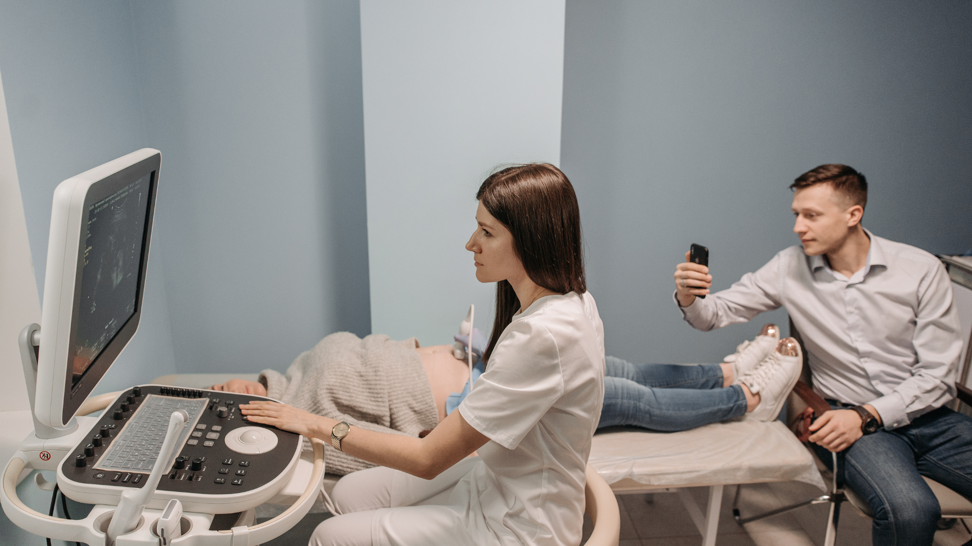
Roles and Expertise of MFM Specialists
1.Manage high-risk pregnancies in women with underlying conditions such as diabetes, hypertension, heart disease, kidney disease, or infections
2.Assess and monitor fetal abnormalities, including chromosomal disorders, fetal growth restriction (FGR), and multiple pregnancies
3.Utilize advanced technologies such as high-resolution ultrasound, Doppler ultrasound, amniocentesis, chorionic villus sampling (CVS), and cordocentesis to diagnose structural abnormalities, genetic conditions, and evaluate fetal health
4.Provide genetic counseling for families at risk of inherited conditions
5.Collaborate with obstetricians, neonatologists, and multidisciplinary teams to manage complex pregnancy conditions
Technologies in Maternal-Fetal Medicine (MFM)
1.High-resolution (Level II) Ultrasound
- Evaluates fetal structural abnormalities (brain, heart, kidneys, spine)
- Assesses fetal health, placenta position, and amniotic fluid
- Commonly performed at 18-22 weeks
2.Doppler Ultrasound
- Measures blood flow in fetal vessels (umbilical cord, brain/MCA)
- Detects fetal hypoxia or anemia
3.Non-Invasive Prenatal Testing (NIPT)
- Analyzes fetal DNA from maternal blood
- Screens for chromosomal abnormalities (e.g., Trisomy 21, 18, 13)
- Safe, no risk of miscarriage
4.Invasive Prenatal Diagnostic Procedures
- CVS (Chorionic Villus Sampling): Placental tissue testing (11–13 weeks)
- Amniocentesis: Amniotic fluid testing (16–20 weeks)
- Cordocentesis: Fetal blood sampling (18–22 weeks)
- Used for diagnosing genetic disorders and chromosomal abnormalities
Note: Slight miscarriage risk (1-2%)
5.Fetal Echocardiography
- Detailed assessment of fetal heart structure and function
- Recommended for high-risk cases (e.g., maternal diabetes, family history)
6.3D/4D Ultrasound
- Provides detailed 3D images and real-time motion
- Helps detect abnormalities such as cleft lip/palate
7.Fetal Monitoring
- Includes NST, BPP, and CTG
- Evaluates fetal well-being, heart rate, and oxygen status
Source : Overbrook Hospital ChiangRai
Arokago Providers Overbrook Hospital ChiangRai
**Translated and compiled by ArokaGO Content Team
Independent Writer
Share this article
More Articles
Discover more insights on health care and medical tourism.

Your Private Path to Timeless Wellness: Why Medical Wellness by Hertitude Clinic
Some of the most personal conversations a person will ever have take place in a doctor's office. A discussion about biological age, family history, hormonal changes, energy, sleep, skin or hair is rarely just clinical. It touches identity, work, relationships and how someone hopes to live in the decades ahead.

What Are Exosomes? What Do They Do in the Body?
Why do small cuts on your skin heal on their own? When an injury occurs, how do the body’s cells know where to begin the repair process?

What Is a Facelift? Exploring Surgery at Asia Cosmetic Hospital
Sagging cheeks, a less defined jawline, and changes in the skin around the neck often lead people to research facelift surgery. Before choosing treatment, it helps to understand that facial aging involves more than the surface of the skin. Different facelift procedures also address different areas.