Why MFM Specialists Matter: What Every Expecting Mother Should Know Before Antenatal Care

Specialists in Maternal-Fetal Medicine (MFM) play a vital role in managing high-risk pregnancies. With advanced technology, structured care planning, and a multidisciplinary approach, they help reduce risks for both mother and baby from pregnancy through delivery.
Importance of Maternal-Fetal Medicine (MFM)
MFM plays a crucial role in helping high-risk pregnant women achieve safer pregnancies and deliveries by focusing on complication prevention, accurate diagnosis, and personalized care planning tailored to each individual.
Roles and Expertise of MFM Specialists
1.Manage high-risk pregnancies in women with underlying conditions such as diabetes, hypertension, heart disease, kidney disease, or infections
2.Assess and monitor fetal abnormalities, including chromosomal disorders, fetal growth restriction (FGR), and multiple pregnancies
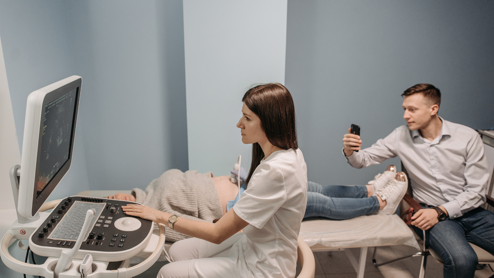
3.Utilize advanced technologies such as high-resolution ultrasound, Doppler ultrasound, amniocentesis, chorionic villus sampling (CVS), and cordocentesis to diagnose structural abnormalities, genetic conditions, and evaluate fetal health
4.Provide genetic counseling for families at risk of inherited conditions
5.Collaborate with obstetricians, neonatologists, and multidisciplinary teams to manage complex pregnancy conditions
Technologies in Maternal-Fetal Medicine (MFM)
1.High-resolution (Level II) Ultrasound
- Evaluates fetal structural abnormalities (brain, heart, kidneys, spine)
- Assesses fetal health, placenta position, and amniotic fluid
- Commonly performed at 18-22 weeks
2.Doppler Ultrasound
- Measures blood flow in fetal vessels (umbilical cord, brain/MCA)
- Detects fetal hypoxia or anemia
3.Non-Invasive Prenatal Testing (NIPT)
- Analyzes fetal DNA from maternal blood
- Screens for chromosomal abnormalities (e.g., Trisomy 21, 18, 13)
- Safe, no risk of miscarriage
4.Invasive Prenatal Diagnostic Procedures
- CVS (Chorionic Villus Sampling): Placental tissue testing (11–13 weeks)
- Amniocentesis: Amniotic fluid testing (16–20 weeks)
- Cordocentesis: Fetal blood sampling (18–22 weeks)
- Used for diagnosing genetic disorders and chromosomal abnormalities
Note: Slight miscarriage risk (1-2%)
5.Fetal Echocardiography
- Detailed assessment of fetal heart structure and function
- Recommended for high-risk cases (e.g., maternal diabetes, family history)
6.3D/4D Ultrasound
- Provides detailed 3D images and real-time motion
- Helps detect abnormalities such as cleft lip/palate
7.Fetal Monitoring
- Includes NST, BPP, and CTG
- Evaluates fetal well-being, heart rate, and oxygen status
Source : Overbrook Hospital ChiangRai
Arokago Providers Overbrook Hospital ChiangRai
**Translated and compiled by ArokaGO Content Team
Independent Writer
Share this article
More Articles
Discover more insights on health care and medical tourism.

5 Food Sources of Vitamin D
We have recently heard a great deal about the importance of vitamin D. On average, the body requires approximately 600-800 IU of vitamin D per day.

How Is Office Syndrome Treated with Thai Traditional Medicine?
“I feel better after a massage, but after a while, the pain comes back again.” This is one of the most common things many office syndrome patients often say. The reason is that relaxation massage ≠ office syndrome treatment. General massage only works on the superficial muscle layer and does not address the root cause of deep-level blockage.

What Causes a Sagging Belly? A Common Concern That Can Be Improved and Made Firm Again
Whether you have a higher body weight or a slim body shape, the problem of a “sagging belly” or loose abdominal skin can equally affect your confidence. Many people try sit-ups, diet control, or intense exercise, but the belly may still appear loose, hanging, or folded downward, without becoming flat, firm, or toned.